Detached and Torn Retina
A Closer Look
What is the Retina?
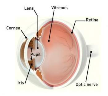
The retina is a nerve layer at the back of your eye that senses light and sends images to your brain.
An eye is like a camera. The lens in the front of the eye focuses light onto the retina. You can think of the retina as the film that lines the back of a camera.
What is Retinal Detachment?
A retinal detachment occurs when the retina is pulled away from its normal position. The retina does not work when it is detached. Vision is blurred, just as a photographic image would be blurry if the film were loose inside the camera.
A retinal detachment is a very serious problem that almost always causes blindness unless it is treated.
What Causes Retinal Detachment?
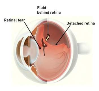
A clear gel called vitreous (vit-ree-us) fills the middle of the eye. As we get older, the vitreous may pull away from its attachment to the retina at the back of the eye.
Usually the vitreous separates from the retina without causing problems. But sometimes the vitreous pulls hard enough to tear the retina in one or more places. Fluid may pass through the retinal tear, lifting the retina off the back of the eye, much as wallpaper can peel off a wall.
The following conditions increase the chance of having a retinal detachment:
- Nearsightedness
- Previous cataract surgery
- Glaucoma
- Severe injury
- Previous retinal detachment in your other eye
- Family history of retinal detachment;
- Family history of retinal detachment
- Weak areas in your retina that can be seen by your ophthalmologist.
What are the Warning Symptoms of Retinal Detachment?
These early symptoms may indicate the presence of a retinal detachment:
- Flashing lights
- New floaters
- A shadow in the periphery (side) of your field of vision
- A gray curtain moving across your field of vision
These symptoms do not always mean a retinal detachment is present; however, you should see your ophthalmologist as soon as possible.
Your ophthalmologist can diagnose retinal detachment during an eye examination and will dilate (enlarge) the pupils of your eyes. Some retinal detachments are found during a routine eye examination.
Only after careful examination can your ophthalmologist tell whether a retinal tear or early retinal detachment is present.
What Treatment is Needed?
Retinal Tears
Most retinal tears need to be treated with laser surgery or cryotherapy (freezing), which seals the retina to the back wall of the eye.
These treatments cause little or no discomfort and may be performed in your ophthalmologist's office. Treatment usually prevents retinal detachment.
Retinal Detachments
Almost all patients with retinal detachments require surgery to return the retina to its proper position.
Types of Surgery
There are several ways to fix a retinal detachment. The decision about which type of surgery and anesthesia (local or general) to use depends upon the characteristics of your detachment.
In each of the following methods, your ophthalmologist will locate the retinal tears and use laser surgery or cryotherapy to seal the tear.
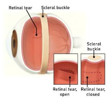
Scleral Buckle
This treatment involves placing a flexible band (scleral buckle) around the eye to counteract the force pulling the retina out of place.
An ophthalmologist often drains the fluid under the detached retina, allowing the retina to settle back into its normal position against the back wall of the eye. This procedure is performed in an operating room.
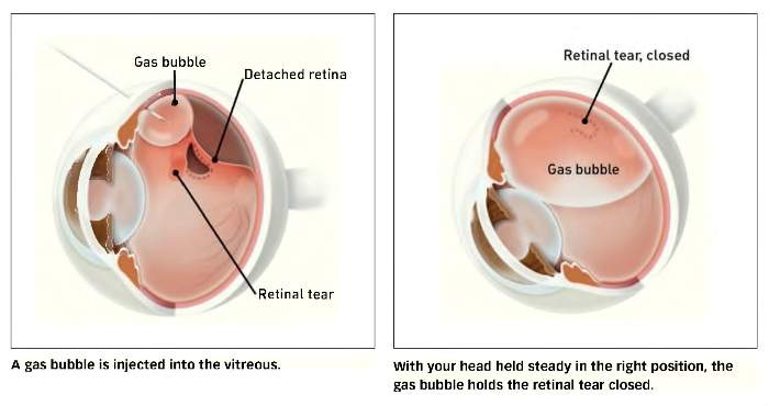
Pneumatic Retinopexy
In this procedure, a gas bubble is injected into the vitreous space inside the eye in combination with laser surgery or cryotherapy. The gas bubble pushes the retinal tear closed against the back wall of the eye. Sometimes this procedure can be done in the ophthalmologist's office.
Your ophthalmologist will ask you to maintain a certain head position for several days. The gas bubble will gradually disappear.
Vitrectomy
This surgery is commonly used to fix a retinal detachment and is performed in an operating room. The vitreous gel, which is pulling on the retina, is removed from the eye and usually replaced with a gas bubble. Sometimes an oil bubble is used (instead of a gas bubble) to keep the retina in place.
Your body's own fluids will gradually replace a gas bubble. An oil bubble will need to be removed from the eye at a later date with another surgical procedure.
Sometimes vitrectomy is combined with a scleral buckle.
After Surgery
You can expect some discomfort after surgery. Your ophthalmologist will prescribe any necessary medications for you and advise you when to resume normal activity. You will need to wear an eye patch for a short time.
Flashing lights and floaters may continue for a while after surgery.
If a gas bubble was placed in your eye, your ophthalmologist may recommend that you keep your head in special positions for a time.
Do not fly in an airplane or travel at high altitudes until you are told the gas bubble is gone! A rapid increase in altitude can cause a dangerous rise in eye pressure. With an oil bubble, it is safe to fly on an airplane.
A change of eyeglasses is often helpful after several months.
What are the Risks of Surgery?
Any surgery has risks; however, an untreated retinal detachment usually results in permanent, severe vision loss or blindness.
Some of the surgical risks include:
- Infection
- Bleeding
- High pressure in the eye
- Cataract
Most retinal detachment surgeries are successful, although a second operation is sometimes needed.
Some retinal detachments cannot be fixed. If the retina cannot be reattached, the eye will continue to lose sight and ultimately become blind.
Will Your Vision Improve?
After successful surgery, vision may take many months to improve and in some cases may never return fully. Unfortunately, some patients do not recover any vision.
The more severe the detachment, the less vision may return. For this reason, it is very important to see your ophthalmologist at the first sign of any trouble.